In the relentless pursuit of modeling complex human neurological and psychiatric diseases, scientists have long turned to "organoids"—three-dimensional structures grown from stem cells that mimic aspects of actual organ architecture. While cerebral organoids have revolutionized in vitro research by recapitulating specialized human cell types and local organizational cues, they have persistently lacked systemic integration. Completely untethered from a vascular system, immune networks, and long-range neural circuits, standard organoids remain isolated islands of tissue, incapable of true functional behavior.
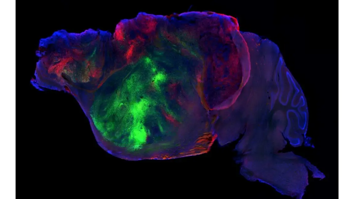
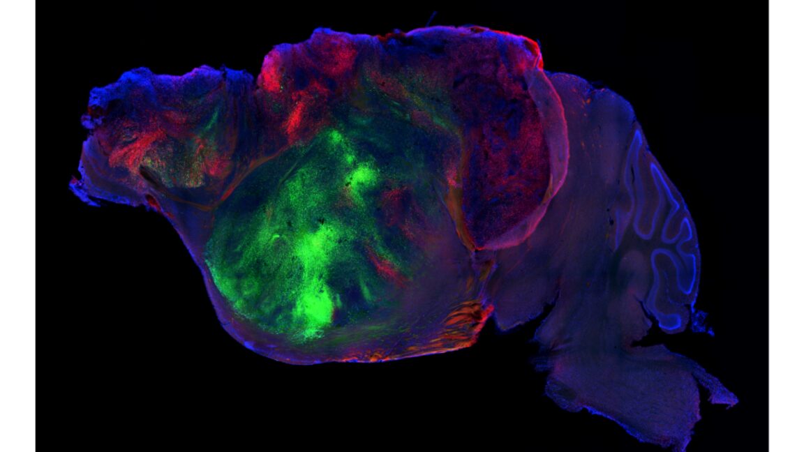
To surmount this physiological isolation, a pioneering research team at Stanford University has executed a radical experimental paradigm: genetically ablating a substantial portion of the developing mouse cortex and replacing it directly with human brain organoid cells. Published in Nature, the study details how human cortical organoids can successfully engraft into a mammalian host, occupying more than 90% of the cortical real estate, extending long-distance axonal projections down to the spinal cord, and generating synchronized neural activity spikes.
However, this structural integration is far from seamless. The resulting "humanized" mice display an organizational structural deficit—notably lacking the distinct laminar layers characteristic of a native mammalian cortex. Behavioral and cognitive assays reveal an ambivalent functional outcome: while the humanized mice perform significantly better than cortex-deficient animals in select spatial and motor evaluations, they fall short of wild-type mice and fail to recover complex associative memory. Consequently, while this breakthrough marks an extraordinary leap forward in chimeric neuroscience, it also underscores the immense biological hurdles that remain before such models can reliably simulate intricate human neurodegenerative conditions like amyotrophic lateral sclerosis (ALS) or Alzheimer’s disease.
Detailed Chronology: Engineering the Chimeric Brain
The journey toward integrating human neural tissues into a living mammalian host required overcoming severe developmental and immunological roadblocks. The Stanford team’s methodological breakthrough unfolded across several meticulously managed stages:
1. Genomic Ablation of the Murine Cortex
The primary barrier to implanting human neural tissue into an animal host is the host’s own neurodevelopmental architecture. Mouse neurons mature exponentially faster than human counterparts, and a developing murine brain relies on precise cell-to-cell signaling during a compressed 21-day gestation. Simply injecting human stem cells into a standard, intact mouse brain results in foreign cells being dominated and structurally sidelined by robust host networks.
To clear a biological niche, the Stanford researchers targeted the cortex—the brain region responsible for high-order processing, decision-making, and sensory integration. Utilizing a genetic strategy, they identified a gene active across almost all nascent cortical cells and manipulated it to trigger the deletion of an essential chromosomal separation gene during cell division. This selectively depleted the vast majority of cells destined to form the mature mouse cortex, effectively halving the brain’s overall volume while leaving the subcortical machinery intact.
2. Neonatal Life Support and Immunosuppression
The collateral damage of wiping out the murine cortex posed immediate survival threats to the neonatal pups. Without intervention, these animals could not compete for resources or manage baseline physiology.
Nutritional Scaffolding: Researchers culled litter sizes to ensure that the cortex-deficient pups received uninterrupted maternal nursing. They extended the nursing window and subsequently transitioned the animals to a high-calorie diet.
Immunological Safeguarding: Because foreign human tissue would naturally trigger a hyper-acute host immune response, the murine hosts were rendered immunocompromised. (Researchers noted that in a sterile laboratory housing facility, this vulnerability is mitigated).
Through these intensive care measures, the team achieved nearly full survival rates among mice lacking a native cortex, setting the stage for transplantation.
3. Organoid Implantation and Engraftment
With a vacant anatomical cavity prepared within the developing brain, human cortical organoids were surgically implanted directly into the depleted region. Over subsequent weeks and months, the tissue vascularized and fused with the surrounding subcortical architecture.
Histological analysis revealed that over 85% of the implanted animals successfully integrated the human graft. Remarkably, the human cells expanded to comprise approximately 92% of the cortical volume in these hosts. Rather than being rejected or encapsulated, the human cells proliferated, adopting the spatial parameters left by the ablated mouse tissue and generating all major neuronal subtypes native to a human cortex.
Supporting Context & Metrics: Structural and Behavioral Realities
To quantify the physiological and cognitive impact of the humanized cortical grafts, the Stanford team deployed a battery of cellular analyses, automated video-tracking systems, and cognitive testing paradigms.
Cellular Integration and Long-Range Connectivity
The structural success of the grafts was evaluated using advanced neuroimaging and electrophysiological tracing:
Axonal Projections: Human neurons did not merely sit in isolation; they successfully extended long-distance axonal processes across anatomical boundaries, with human signals detected as far away as the spinal cord.
Electrophysiological Synchronization: Multi-electrode recordings confirmed that the human neurons engaged in synchronized electrical activity spikes, indicating local network coordination.
Laminar Deficits: Despite local cell-type clustering (where cells that typically reside in specific cortical layers tended to aggregate near one another), the hallmark laminar architecture—the distinct multi-layered striping of a healthy cortex—completely failed to form.
Behavioral and Cognitive Phenotyping
To determine whether this humanized architecture translated into functional recovery, researchers evaluated the mice across three distinct cohorts: normal (wild-type) mice, cortex-deficient mice, and humanized-cortex chimeras.
Behavioral/Cognitive Metric
Cortex-Deficient Mice
Humanized-Cortex Mice
Wild-Type (Normal) Mice
Locomotion & Behavior Clusters
Distinct, erratic patterns
Formed a distinct third cluster
Baseline normal patterns
Body Weight Trajectory
Significantly underweight
Intermediate weight
Baseline normal weight
Spatial Maze Memory
Performance equal to random chance
Moderate improvement over chance
High success rate
Associative Memory Tests
No better than chance
No better than cortex-deficient mice
High success rate
Fine Motor Coordination
Severe impairment
Intermediate recovery
Baseline normal recovery
The data paints a nuanced picture: having a disorganized human replacement cortex provided a measurable functional upgrade over having no cortex at all, yet it remained vastly inferior to an intact, native mammalian brain.
Official Statements and Expert Perspectives
The publication of this study in Nature has ignited widespread debate across the global neuroethics and neurobiology communities. While the technical achievement of substituting a major mammalian brain structure with human tissue is universally acknowledged as a monumental feat of bioengineering, experts urge caution regarding its immediate translational utility.
"The results are only a slight improvement over missing the entire brain structure," notes the lead research documentation from the S. Pasca lab at Stanford University, highlighting the modest cognitive gains observed in the chimeric subjects.
Independent neuroscientists reviewing the data have emphasized that the absence of structured laminar organization poses a fundamental roadblock. Without the precise circuitry found in natural brains, modeling complex human-specific disorders—such as schizophrenia, autism spectrum disorders, or severe neurodegenerative cascades—remains an aspiration rather than a current reality.
Furthermore, the ethical dimensions of creating mammalian brains embedded with significant proportions of human neural tissue continue to draw scrutiny from institutional review boards and bioethicists alike. Although current cognitive metrics suggest the mice are far from achieving human-like consciousness or higher-order reasoning, the rapid trajectory of chimeric research necessitates stringent, proactive regulatory frameworks.
Future Outlook: The Path Ahead for Chimeric Neuroscience
As the dust settles on this initial proof-of-concept study, the scientific community faces critical technical hurdles before human-mouse chimeras can transition from experimental novelties to indispensable disease-modeling tools.
1. Decoding Cellular Micro-Environments
Future iterations of this research must focus on deciphering why human organoids fail to self-organize into proper laminar layers when placed inside a murine host. Researchers hypothesize that biochemical cues from the host environment—or the absence of specific human-specific extracellular matrix signals—may be missing. Identifying and supplementing these missing developmental signposts will be essential for engineering structurally faithful tissue.
2. Moving Beyond Hypoxia Studies
To date, the primary validated functional response of these humanized grafts has been their distinct physiological reaction to brief periods of hypoxia (oxygen deprivation), which mirrors normal human cellular thresholds. While this confirms that the human cells are metabolically active and responsive, it falls short of modeling polygenic, systemic neural pathologies like ALS or Alzheimer’s. Expanding the functional repertoire of the graft to model synaptic pruning, neuroinflammation, and protein aggregation will be the definitive test of the model’s utility.
3. Ethical Governance and Transparency
As chimeric models grow increasingly sophisticated, transparency in reporting cognitive advancements and pain-sensing capabilities will remain paramount. Institutional oversight committees will need to continuously re-evaluate the moral status of animals harboring human neural circuitry, ensuring that scientific ambition is balanced with rigorous ethical stewardship.
In summary, Stanford’s partial cortical replacement model represents a daring step into uncharted biological territory. By demonstrating that human neural tissue can physically integrate, wire into spinal pathways, and partially rescue the deficits of an ablated brain, the study shatters previous boundaries of what is possible in regenerative biology. Yet, as the mice’s intermediate cognitive scores remind us, building a mind is infinitely more complex than simply swapping its parts.