








Executive Overview
For generations, sleep research has viewed the cerebral cortex as a passive passenger—a high-level processing organ that merely reacts to deep-seated subcortical directives from the brainstem and hypothalamus. The prevailing neuroscientific dogma held that while the slow rhythms characteristic of deep, restorative sleep are clearly observable in the cortex, the actual master signals triggering them must originate elsewhere.
A groundbreaking study recently published in Nature shatters this long-standing assumption. Led by researchers Geoffrey Terral and Renata Batista-Brito at the Albert Einstein College of Medicine in New York, the research team has identified a previously unheralded population of cortical cells that can trigger sleep entirely on their own. These rare cells—designated as Sst-Chodl neurons—account for roughly one percent of the cortex’s inhibitory neurons, translating to a staggering rarity of about one in every thousand cortical cells.
Despite their microscopic demographic footprint, these neurons wield extraordinary macro-level power. When experimentally switched on in mice, these cells rapidly transition the animals into deep slow-wave and REM sleep, bypassing circadian timing entirely to induce slumber even during peak waking hours. This discovery not only upends textbook neuroscience regarding how sleep is initiated, but it also provides a tantalizing entry point into understanding and treating the pervasive sleep disruptions associated with numerous psychiatric and neurological disorders.
Detailed Chronology: The High-Risk Quest for the 1%
The road to discovering Sst-Chodl neurons was defined by high-stakes scientific gambling, career-threatening grant rejections, and years of methodical genetic troubleshooting.
Overcoming the "Contaminant" Problem
Long-range inhibitory neurons marked by specific genetic signatures have been studied in primates for years. However, neuroscientists continually hit a brick wall when attempting to isolate and manipulate them. Targeting either of the two active genes within these cells—Sst or Chodl—inevitably captured a massive, highly heterogeneous family of diverse neurons.
"A single gene is not able to target these cells," explains Batista-Brito. Because inhibitory neurons make up roughly 20% of all cortical neurons, isolating a subset that represents just 0.1% of the total population meant that any broad genetic approach would introduce overwhelming "noise." As Batista-Brito notes, "If you’re not really specific, the contaminants are going to be much more dominant than the specific cells."
Building a genetic strategy capable of labeling a cell only when both genes were simultaneously active required years of meticulous trial and error. Funding agencies were deeply skeptical. "Going after these cells was really a high-risk project because the likelihood of seeing anything with 0.1 percent of neurons in the cortex is really low," Batista-Brito recalls. "I wrote a bunch of grants on these projects that were always rejected because it was too high-risk."
Unveiling Massive Arborization
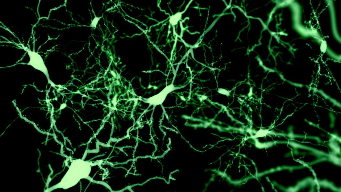
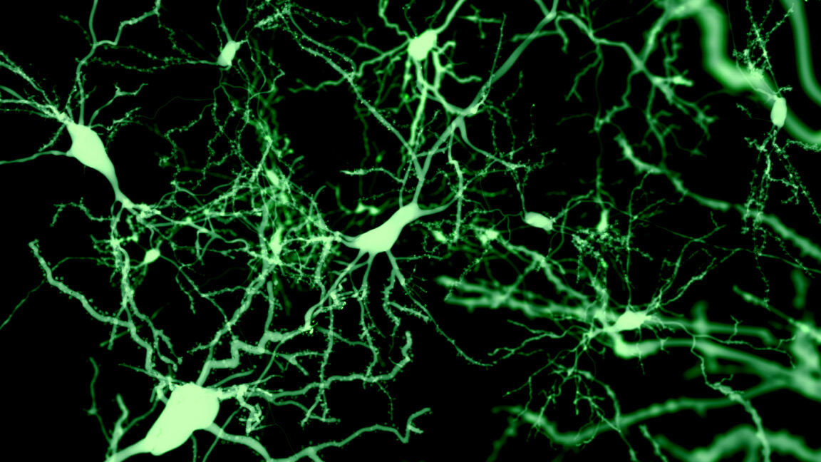
The gamble finally paid off when the team visualized the anatomy of the successfully labeled Sst-Chodl neurons. The initial view under the microscope revealed a stunning paradox: "At first, I saw two or three cell bodies in the whole brain," Batista-Brito says. "Despite that, there was massive, massive arborization all over the visual cortex, like I never saw with any other neuron."
This extraordinary "arborization"—a tree-like branching structure connecting neurons across vast neuro-anatomical distances—flies in the face of classical neuroanatomy. Typical inhibitory neurons are local operators; they receive inputs from various brain regions and exert control exclusively over their immediate, microscopic patch of tissue.
Sst-Chodl neurons do precisely the opposite. They receive remarkably precise, localized inputs, but then broadcast that information globally. A single Sst-Chodl cell branches extensively across the entire visual cortex while projecting long axons to brain regions dedicated to touch, hearing, spatial memory, navigation, and voluntary movement.
Supporting Context & Metrics: Decoding the Neural Mechanics
To understand how these hyper-connected cells operate during natural sleep cycles, Terral and Batista-Brito deployed simultaneous multi-modal imaging. They tracked neuronal firing alongside physiological markers in mice, including pupil size, muscle tone, facial micro-movements, running behavior, and cortical electrical activity.
The Quiet Hours and Breaking the UP/DOWN Pattern
Of the 111 Sst-Chodl cells imaged during the experiment, an overwhelming 95 lit up precisely during slow-wave sleep and quiet, motionless wakefulness. Conversely, they fell completely silent during periods of active running and REM sleep.
During slow-wave sleep, the healthy cerebral cortex naturally alternates between "UP states" (periods of vigorous, synchronized firing) and "DOWN states" (periods of near-total silence). Sst-Chodl neurons shatter this rhythmic status quo.
- Peak Firing at Termination: "Their activity is even higher at the termination of the UP state," notes Terral. Just as surrounding neurons begin to wind down their activity, Sst-Chodl cells spike even higher.
- Skipping the Rebound: While other neurons show a predictable electrical "rebound" when emerging from a DOWN state, Sst-Chodl cells bypass this entirely. Their unique electrophysiological profile sets them apart from every other measured cell population during state transitions.
Optogenetic Control and Delta Wave Generation
To move from observation to causation, the team utilized optogenetics—introducing light-activated ion channels into the Sst-Chodl neurons so they could be fired artificially on command using targeted pulses of light.
When stimulated optogenetically in the visual cortex, the results were profound:
- Delta Power Amplification: The stimulation drove delta power—the slowest, highest-amplitude brain waves that define deep, restorative slow-wave sleep—uniformly across every cortical layer.
- Tighter Spike Timing: The manipulation increased the frequency and duration of DOWN states while keeping overall firing rates nearly static.
- Coordination Over Volume: "Those neurons are not changing so much the firing rate," Terral points out. "They change just the coordination—whether the neurons fire together or not."
Strikingly, the network’s oscillation frequency remained constant regardless of the stimulus pattern applied. Whether researchers delivered a flat stimulus, a delta-frequency pulse, or high-frequency 20Hz or 60Hz bursts, the network invariably slipped into the same signature oscillation. The intrinsic biophysical properties of Sst-Chodl cells dictate that once triggered, they lock into a fixed rhythmic output.
Furthermore, their sphere of influence is immense. Although their cell bodies sit firmly in the visual cortex, direct inhibitory currents were recorded in a third of cells located two full millimeters away, with measurable downstream effects cascading all the way into frontal motor areas.
Official Statements & Expert Insights
The implications of the study extend far beyond basic neuroanatomy, pointing directly toward the brain’s homeostatic sleep regulation systems.
Commenting on the behavioral outcomes, Batista-Brito highlights the unexpected potency of their experiments. When the team stimulated Sst-Chodl neurons across the cortex of freely moving mice, the animals rapidly entered deep slow-wave and REM sleep, exhibited a drastic reduction in sleep-onset latency, and retreated to their nests during daylight hours.
To test the absolute limits of this neural switch, the researchers repeated the stimulation during the dark phase—the natural nocturnal window when mice are normally active and awake.

"We could have them sleep more during the time that they are awake than they usually sleep during the day," Renata Batista-Brito stated. "I never thought this experiment was going to work. We are manipulating one percent of inhibitory neurons with local injections, so we are hitting only a fraction of those cells. And we could see an effect that was really quite striking."
This striking efficacy lends heavy credence to a hypothesis first advanced by co-author Thomas Kilduff, director of SRI International’s Center for Neuroscience. Kilduff proposes that Sst-Chodl neurons act as the cortex’s primary sensors of sleep pressure—the homeostatic fatigue that steadily accumulates the longer an organism stays awake, eventually forcing sleep regardless of circadian signals. Kilduff’s prior work demonstrated that following sleep deprivation, Sst-Chodl cells become the single most active neuronal population in the cortex.
Future Outlook & Next Steps
While the discovery of Sst-Chodl neurons marks a watershed moment in sleep science, several critical mysteries remain to be solved.
1. What Flips the Switch?
The most pressing unanswered question is what natural physiological signals trigger these cells to fire in the first place. Because the initial study focused exclusively on the visual cortex, the team is currently replicating their anatomical mapping work within the prefrontal cortex. Their working hypothesis is that prefrontal Sst-Chodl populations receive direct inputs from deep subcortical structures like the hypothalamus and thalamus, serving as the biological bridge between global metabolic sleep pressure and cortical slow-wave generation.
2. Conservation Across Species
Evolutionary biology suggests this mechanism is ancient and vital. Sst-Chodl neurons are evolutionarily conserved across a vast phylogenetic tree, appearing in species ranging from salamanders to humans. This deep conservation underscores why researchers believe these cells could serve as a precise therapeutic entry point for treating severe sleep dysregulations common to numerous psychiatric and neurodegenerative conditions, including major depression, schizophrenia, and Alzheimer’s disease.
3. The Path Forward
Terral, Batista-Brito, and their colleagues at the Albert Einstein College of Medicine are now dedicating their research pipeline to answering three defining questions:
- What endogenous biological signals activate these cells?
- Do they definitively function as the brain’s internal barometer for sleep pressure?
- By what precise biophysical mechanism do they universally drive high-amplitude delta power across disparate brain networks?
As laboratories around the world begin to investigate these master cortical switches, the long-held view of the cerebral cortex as a passive follower of the deep brain is officially closed. The cortex, it turns out, possesses its own master conductor for sleep—hiding quietly among just one percent of its cells.
Published in Nature (2026). DOI: 10.1038/s41586-026-10876-y