









Executive Overview
In the realm of modern neurology, sudden unilateral tremors are typically flagged as precursors to transient ischemic attacks, acute cerebrovascular events, or localized epileptic seizures. However, an extraordinary clinical case documented in the New England Journal of Medicine has underscored the alarming and unpredictable manifestations of neglected tropical diseases. A female rice farmer from the Philippines experienced a terrifying episode where the entire right side of her body began shaking uncontrollably, entirely unprompted. Displaying no pre-existing headaches, acute confusion, or localized motor weakness in the moments leading up to the incident, the patient presented a confounding diagnostic puzzle to emergency medical personnel.
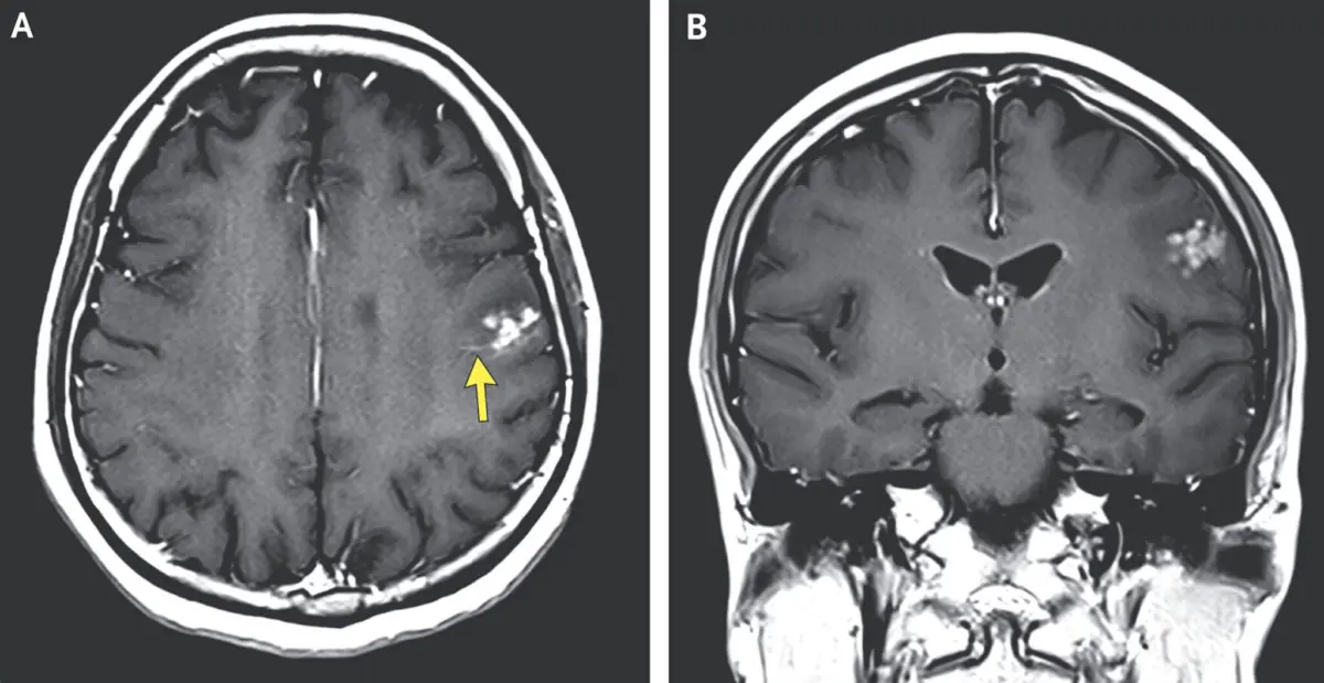
Upon admission and following an unremarkable routine physical examination, neuroimaging unlocked the shocking root cause of her ordeal. Magnetic resonance imaging (MRI) of her brain revealed a distinct, bright cluster of nodules encapsulated by fluid within her left frontal lobe. The medical consensus was as startling as it was rare: the patient’s brain was hosting clusters of parasitic worm eggs.
Doctors ultimately diagnosed the woman with neuroschistosomiasis, a severe and uncommon neurological complication stemming from an infection by Schistosoma japonicum, a parasitic blood fluke. This comprehensive investigative report explores the labyrinthine biological lifecycle of the parasite, the intricate mechanisms of neuroschistosomiasis, the socioeconomic vulnerabilities tied to agricultural labor, and the broader implications for global public health surveillance in an increasingly interconnected world.
Detailed Chronology: From Farm to Emergency Room
The Sudden Onset
The clinical timeline began without warning. While performing routine daily activities, the patient—whose livelihood as a rice farmer exposes her daily to standing water systems endemic to Southeast Asia—felt a strange, rhythmic juddering begin in her right arm. Within seconds, the violent, involuntary tremors spread downward to her right leg.
For approximately sixty agonizing seconds, the right half of her body shook uncontrollably. Remarkably, the episode subsided as abruptly as it had materialized. Unlike patients experiencing classic focal seizures or acute strokes, she retained complete lucidity. She experienced no post-ictal confusion, no throbbing cephalalgia, and no residual motor deficits once the shaking ceased. Recognizing the alarming nature of the event, she immediately sought medical evaluation at a local emergency department.
Clinical Assessment and Diagnostic Imaging
Upon her arrival at the hospital, an attending medical team performed a comprehensive physical and neurological examination. To the surprise of the clinicians, the preliminary physical exam yielded entirely normal results. Her cranial nerves were intact, deep tendon reflexes were symmetric, and coordination appeared uncompromised.
Had the diagnostic protocol ended with the physical exam, the underlying pathology may have been entirely missed, leaving the patient vulnerable to recurrent, potentially catastrophic neurological events. Recognizing the necessity of evaluating the central nervous system following unexplained paroxysmal motor episodes, the medical team ordered an advanced neuroimaging session.
The MRI scan provided the definitive turning point. Detailed cross-sectional imagery of the brain uncovered a conspicuous cluster of hyperintense nodules surrounded by localized edema (fluid accumulation) residing squarely within the left frontal lobe—the region responsible for voluntary motor control on the right side of the human body. Based on the morphological presentation of the nodules and the patient’s occupational history, the diagnostic team immediately suspected parasitic ova deposition within cerebral tissues.
Confirmation and Treatment
Subsequent case analyses published in the New England Journal of Medicine confirmed the clinical suspicion: neuroschistosomiasis. This condition arises when Schistosoma eggs bypass the traditional hepato-intestinal venous system, aberrantly migrating through the venous plexus of Batson or via arterial shunts into the central nervous system.
Management of such cases requires a delicate balance of antiparasitic pharmacotherapy—typically praziquantel—to eradicate the adult flukes, coupled with targeted corticosteroid regimens to dampen the severe localized inflammatory and granulomatous immune responses triggered by the trapped eggs within the neural parenchyma. For this Filipino rice farmer, the prompt identification of the nodules prevented long-term neurological sequelae, illustrating the critical importance of maintaining a broad differential diagnosis that incorporates geographical and occupational risk factors.
Supporting Context & Metrics: The Diabolical Lifecycle of Schistosoma japonicum
To comprehend how a microscopic organism residing in a rural Philippine paddy could infiltrate the human central nervous system, one must examine the remarkably complex, multi-host lifecycle of the blood fluke. Schistosoma japonicum belongs to a genus of trematodes responsible for schistosomiasis, a chronic disease impacting hundreds of millions of people globally, predominantly in tropical and subtropical developing regions.
[Human / Animal Host] ---> (Eggs in Feces/Urine) ---> [Freshwater Environment]
         ^                                                      |
         |                                           (Ciliated Miracidia Hatch)
         |                                                      v
[Cercariae Penetrate Skin] <--- (Asexual Reproduction) <--- [Aquatic Snail]
Phase One: Environmental Shedding and Miracidia
The lifecycle begins when infected mammalian hosts—including humans, water buffalo, rodents, and pigs—shed parasite eggs via their feces or urine into freshwater ecosystems. When these waste products contaminate rural water supplies, the eggs encounter freshwater environments and hatch, releasing free-swimming, ciliated larvae known as miracidia.
Phase Two: Intermediate Snail Hosts
Driven by chemical cues, the microscopic miracidia actively hunt for specific species of amphibious or aquatic freshwater snails (Oncomelania species, in the case of S. japonicum). Upon locating a suitable host, the larvae bore directly into the snail’s soft tissues.
Inside the snail, the parasite undergoes extensive asexual multiplication and development, transforming through two distinct larval stages (sporocysts and rediae). This biological amplification converts a single miracidium into thousands of infectious offspring.
Phase Three: Cercariae and Skin Penetration
Following weeks of development, thousands of late-stage larvae—now equipped with characteristic forked tails and referred to as cercariae—burst out of the host snail and return to the water column. These free-swimming cercariae are remarkably resilient and possess specialized proteolytic glands that enable them to detect human skin chemistries.
When an agricultural worker, such as a rice farmer, wades barefoot or barehanded into flooded fields, the cercariae swarm the exposed skin. Utilizing energetic muscular movements and enzymatic secretions, these microscopic invaders tunnel directly through intact human skin, shedding their forked tails in the process.
Phase Four: Systemic Migration and Pairing
Once inside the human vascular system, the transformed larvae—now called schistosomules—embark on an internal odyssey. They travel via the peripheral venous circulation to the heart and lungs, subsequently navigating the pulmonary capillary beds to reach the hepatic portal system (the liver).
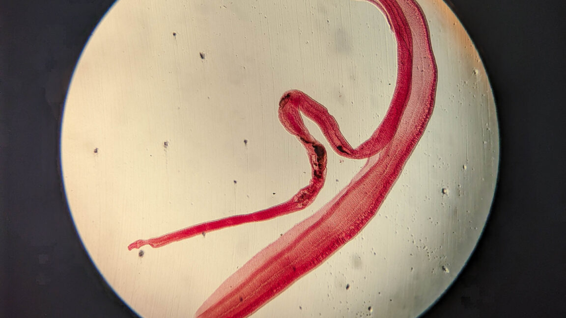
In the liver, the parasites mature into adult male and female worms over several weeks. Once mature, the adult worms pair up in a permanent embrace: the slender female worm tucks snugly into a specialized gynecophoral canal running along the length of the thicker male worm.
These permanent couples migrate to their final ecological niche within the large mesenteric blood vessels surrounding the intestines or urinary bladder. Here, the female deposits hundreds to thousands of eggs daily. While many eggs successfully penetrate the intestinal wall to exit the body and perpetuate the cycle, a fraction become trapped in local tissues—or, in anomalous cases, drift upstream into cerebral or spinal tissues, causing severe ectopic pathologies like neuroschistosomiasis.
Official Statements & Epidemiological Perspectives
Global health organizations, including the World Health Organization (WHO) and the Centers for Disease Control and Prevention (CDC), monitor schistosomiasis as one of the most significant Neglected Tropical Diseases (NTDs) affecting rural agricultural communities.
Geographic Distribution and Elimination Milestones
Schistosoma japonicum exhibits a distinct geographical footprint across East and Southeast Asia. While historical endemicity was widespread, rigorous public health interventions over the decades have yielded major milestones. Notably, through massive deworming campaigns, environmental management, and improved sanitation, S. japonicum has been officially eliminated from Japan.
However, the parasite remains stubbornly entrenched in parts of the People’s Republic of China, select regions of Indonesia, and various agricultural provinces throughout the Philippines. In these areas, reliance on manual farming techniques—particularly wet-rice cultivation—keeps rural populations in continuous contact with contaminated water systems.
Clinical Insights from Parasitologists
Infectious disease specialists emphasize that neuroschistosomiasis remains an underdiagnosed clinical entity. Because cerebral schistosomiasis mimics other neurological disorders—ranging from primary brain tumors and tuberculomas to conventional cerebral toxoplasmosis and high-grade gliomas—patients frequently undergo unnecessary surgical biopsies or prolonged, ineffective treatments before advanced imaging and detailed occupational histories reveal the true parasitic etiology.
Medical toxicologists and parasitologists publishing in the New England Journal of Medicine highlight the necessity of localized epidemiological vigilance:
"When clinicians encounter atypical focal neurological presentations—such as sudden, unexplained motor tremors accompanied by localized brain lesions—in patients with histories of occupational exposure in endemic freshwater zones, parasitic infections must remain prominent in the differential diagnostic framework."
Future Outlook: Mitigation, Diagnostics, and Control
Addressing the ongoing threat of neuroschistosomiasis and reducing the disease burden of blood flukes requires a multifaceted, multidisciplinary approach spanning clinical medicine, veterinary science, environmental engineering, and public policy.
Advancements in Diagnostic Sensitivity
Early detection remains the cornerstone of preventing severe neurological complications. Traditional diagnostic methods rely heavily on microscopic stool examinations (Kato-Katz thick smear technique) or serological antibody assays. However, stool microscopy often lacks sensitivity in low-intensity infections or ectopic presentations like neuroschistosomiasis, where adult worms reside outside the gastrointestinal tract and shed few or no eggs into feces.
Future clinical protocols are increasingly shifting toward sensitive molecular diagnostics, such as polymerase chain reaction (PCR) assays targeting circulating parasite DNA or circulating cathodic antigen (CCA) detection tests. These tools allow for rapid, non-invasive identification of active infections long before catastrophic granulomatous reactions manifest in the central nervous system.
Environmental Engineering and One Health Strategies
Because S. japonicum relies heavily on mammalian reservoir hosts beyond humans—such as water buffalo, cattle, goats, and domestic rodents—controlling the parasite solely through human mass drug administration (MDA) with praziquantel is insufficient.
Public health strategists are embracing the "One Health" paradigm, which recognizes that human health is inextricably linked to animal health and the shared environment. Control frameworks moving forward must incorporate:
- Veterinary Interventions: Treating livestock reservoir hosts to block environmental egg contamination.
- Snail Control: Targeted molluscicide application and environmental habitat modification (such as concrete-lining irrigation canals) to suppress Oncomelania snail populations.
- Infrastructure Development: Expanding access to clean, treated municipal water supplies and modern sanitation facilities in rural agricultural zones to break the transmission cycle at its source.
Empowering Vulnerable Workforces
Ultimately, safeguarding agricultural workers like the Filipino rice farmer featured in this landmark case requires systemic socioeconomic investments. Providing personal protective equipment—such as rubberized boots and gloves—combined with community-wide health education campaigns regarding the risks of occupational exposure to stagnant agricultural waters, will empower vulnerable populations to protect themselves.
As climate patterns shift and agricultural practices evolve, maintaining robust international surveillance systems ensures that rare and complex tropical infections like neuroschistosomiasis are identified swiftly, treated effectively, and understood comprehensively within the broader landscape of global medicine.