








Executive Overview
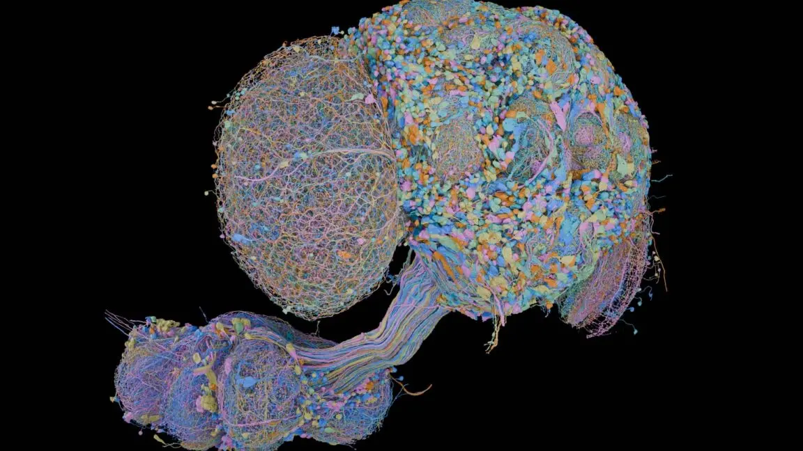
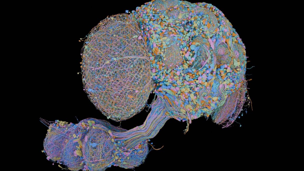
In a milestone that bridges the gap between high-throughput computational engineering and cellular neurobiology, researchers have officially announced the completion of a comprehensive, wire-by-wire connectome of the male fruit fly (Drosophila melanogaster) brain. Published in Cell, the map details every single neuron, spatial location, and intercellular contact within a male fly’s central nervous system—revealing a staggering network of roughly 150,000 neurons bridged by more than 300 million individual synaptic connections.
This achievement is not isolated. Coming on the heels of a female Drosophila connectome completed earlier this year by an independent academic team, the male map establishes a dual-sex baseline for the simplest yet functionally rich model organism in neurobiology. More importantly, the project serves as a monumental proof-of-concept. It demonstrates that mapping the trillions of synapses in vertebrate brains—including mice and, eventually, humans—is an attainable engineering target, provided that biological expertise and cutting-edge artificial intelligence continue their successful alliance.
Detailed Chronology: From Photographic Prints to Generative AI
To appreciate the technological leap required to map 300 million synapses, one must look back at the origins of connectomics. In 1971, Gerry Rubin—now a senior group leader at the Howard Hughes Medical Institute’s (HHMI) Janelia Research Campus and a senior author on the new paper—arrived as a graduate student at the UK’s Laboratory of Molecular Biology. At the time, researchers had acquired a massive computer with ambitions to automate the mapping of Caenorhabditis elegans, a transparent nematode with a modest 300 neurons.
"It took them about two years to realize that the computers were nowhere near powerful enough," Rubin recalls.
Lacking the computational horsepower to process digital imagery, the early pioneers of connectomics abandoned automation. They resorted to printing electron micrographs on physical photographic paper, highlighting neurons with colored magic markers, and manually tracing circuits by hand.
That labor-intensive method hit a hard evolutionary wall when applied to organisms more complex than a microscopic worm. The fruit fly is opaque, rendering its nervous system notoriously difficult to image, and boasts a cell count five hundred times greater than C. elegans.
Breaking this barrier required a four-year interdisciplinary campaign uniting Janelia’s expert biologists with Google Research’s elite computer scientists. The workflow required transforming a physical, dissected male fruit fly brain—along with a portion of its ventral nerve cord—into a digital artifact:
- Physical Sectioning: The tissue was embedded, cured, and sliced into an immense series of ultra-thin, evenly spaced sections.
- Electron Microscopy: Each slice was imaged using high-resolution electron microscopy, capturing the minute ultrastructure of membranes and organelles.
- Computational Stitching & Generative AI: Mechanical slicing invariably introduces physical tears, tissue distortions, and lost material. Google Research developed specialized generative AI models to virtually stitch these blocks together, smoothing out seams and eliminating gaps so that downstream algorithms could process the data uninterrupted.
- Cellular Segmentation: A recurrent visual neural model traversed the voxel data, converting raw microscopic pixels into explicit, three-dimensional spatial outlines of individual neurons.
- Synapse Classification: Additional machine learning models identified and classified synapses, continually refined through an iterative feedback loop between algorithms and human proofreaders.
Supporting Context & Metrics: Decoding the Fly Connectome
The fruit fly brain is a marvel of biological miniaturization. While its entire nervous system contains roughly 150,000 neurons, the brain proper houses only a fraction of that number. Yet, within this compact volume, the automated pipeline detected over 300 million synaptic connections.
To make sense of this intricate wiring diagram, neurobiologists must analyze how sensory inputs are transformed into motor outputs, memories, and behaviors. Sensory neurons register environmental stimuli—light, sound, and touch—passing this data to local processing centers. From there, complex routing determines whether the information triggers an immediate reflex or is shuttled to higher-order cognitive processing areas.
Sex on the Brain: Unpacking the dsx and fru Genes
One of the most profound scientific yields of having both male and female Drosophila connectomes is the ability to map anatomical sexual dimorphism down to individual synapses. Decades of genetic research have established that sex determination in flies is largely driven by two master-regulatory genes: doublesex (dsx) and fruitless (fru). These genes translate the fly’s chromosomal makeup (XX vs. XY) into overt physiological and behavioral differences.
Comparing the male and female connectomes revealed:
- 289 male-specific neurons
- 71 female-specific neurons
- 138 shared neurons that exhibit radically different morphologies and connection topologies between the sexes
The relationship between these structural differences and genetic drivers is remarkably nuanced. Approximately 90% of the male-specific neurons actively expressed dsx and fru. However, 10% of these male-specific cells lacked active expression of both genes, suggesting they acquired their sex-specific identities downstream through intercellular signaling.
Similarly, nearly 40% of the neurons present in both sexes with distinct structural dimorphisms lacked active dsx and fru expression. Conversely, about 7% of identical-looking neurons did express these genes, implying functional divergence independent of physical shape.
Furthermore, the data unveiled unexpected architectural anomalies. For example, two distinct neuron types responsible for controlling a female’s physical mating response were also discovered in males—possessing the exact same basic morphology despite the complete absence of the corresponding female anatomy. Instead of driving absent physical machinery, these shared neurons were re-routed, forming entirely different synaptic connections in the male brain.
By plotting the spatial distribution of these sex-specific cells, researchers uncovered a broad organizational rule: sex differences are generally insulated from raw sensory input and basic motor interfaces. Instead, they cluster within higher-order integrative and decision-making centers, effectively modulating how the brain interprets the world rather than altering how it senses it.
Official Statements and Collaborative Vision
The success of the male fly connectome project underscores a paradigm shift in modern bioscience: the absolute necessity of cross-disciplinary partnerships.
"The reason I think Google and we were interested in this is because this is this type of grand challenge that just cannot be done in any other way," explains Michal Januszewski, a staff scientist at Google Research. "We knew we need AI for this. This cannot be solved by having more humans or by any other technology. And it’s important."
Neither biologists nor computer scientists could have crossed the finish line alone. Biologists provided the foundational wet-lab chemistry, dissection mastery, and anatomical ground-truth validation. Computer scientists supplied the scalable architecture required to process petabytes of microscopic imagery.

Reflecting on the human resource economics of the endeavor, Gerry Rubin emphasizes that scalability is the ultimate metric of success for future connectomics projects:
"Our view is we did Drosophila with a team of 50 people. The hope is, by the time someone does a mouse, they’ll also need a team of 50 people, even though there are a thousand times more neurons in there. The people will never go away, but the people will not need to scale with the number of neurons, which would be economically not feasible."
Future Outlook: Accelerating Neuroscience and Theoretical Horizons
The publication of the male and female Drosophila connectomes marks not an end, but a beginning. Just as the sequencing of the fly genome catalyzed decades of genetic discovery by turning gene identification into a database query, the connectome instantly transforms structural neurobiology. Today, when researchers identify an interesting neuron in vivo, they can open an interactive browser, trace its axonal tree, and map its downstream synaptic targets in minutes rather than years.
Beyond expediting empirical experiments, the connectome is reshaping theoretical neuroscience. Historically, computational theorists operated without definitive wiring constraints, forced to hypothesize abstract models of how a brain might function.
"Before this, most neuro theorists were very much like, ‘How could a brain work?’ And they didn’t have a constraint," Rubin notes. "Once they had the connectome, they could say, ‘The brain does this and here’s the wiring diagram. How can this wiring diagram allow this function?’ So this has been a major… the biggest change in having the connectome."
However, realizing the full statistical and evolutionary potential of connectomics will require overcoming yet another logistical hurdle: throughput.
Among the most intriguing anomalies discovered in the new map was a specific neuron present in the female connectome that was entirely missing from one hemisphere of the male fly’s brain—likely the casualty of a developmental stochastic error. With only two complete adult fly connectomes in existence, science lacks the statistical baseline required to quantify how common such individual variations truly are.
To achieve statistical confidence regarding neural wiring variability, researchers will need to sequence dozens of individual brains. If each connectome continues to require four years of sustained effort, that goal remains out of reach. The ultimate legacy of the male Drosophila project, therefore, lies in the automated pipelines and AI algorithms forged during its creation. As these techniques are refined, automated, and scaled, biology moves ever closer to its ultimate cartographic ambition: charting the complete neural architecture of the mammalian brain.