









Executive Overview
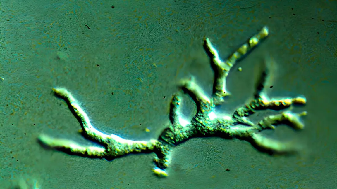
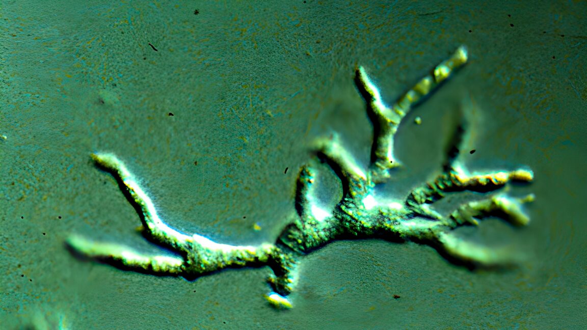
In the realm of modern pediatrics, few nightmares rival the silent, swift progression of a neglected pathogen capable of outsmarting specialized medical teams. A harrowing case study published in BMJ Case Reports by clinicians at Seattle Children’s Hospital sheds light on the terrifying clinical reality of Balamuthia mandrillaris—a free-living, single-celled amoeba found naturally in soil and water. The report details the tragic case of a healthy toddler from Washington state who fell victim to an ultra-rare, almost universally fatal brain infection known as granulomatous amoebic encephalitis (GAE).
What makes this case profoundly disturbing is not merely the devastating outcome, but the winding diagnostic labyrinth that preceded it. Because B. mandrillaris mimics other neuro-inflammatory disorders, initial tests led medical professionals down an entirely erroneous path. Believing the boy suffered from a rare form of systemic vasculitis known as childhood Takayasu arteritis, doctors administered powerful immunosuppressive medications. Far from helping, this intervention likely lowered the child’s defenses, allowing the opportunistic pathogen to colonize and ravage his brain tissue unchecked.
It took more than 30 days of escalating symptoms, false leads, and deteriorating health for clinicians to definitively identify the amoeba via metagenomic next-generation sequencing and brain biopsy. By the time the correct microscopic culprit was exposed, irreversible damage had already stripped the young boy of higher neurological function. His heartbreaking passing, coupled with his parents’ selfless decision to publish the case, now offers the global medical community an invaluable roadmap. By examining where diagnosis failed, infectious disease experts hope to rewrite the diagnostic playbook for future encounters with this invisible, opportunistic killer.
Detailed Chronology: A Month-Long Diagnostic Odyssey
The tragedy unfolded with deceptive suddenness. Initially, the toddler presented as a healthy, vibrant child before abruptly developing an alarming constellation of symptoms: persistent vomiting, profound lethargy, and noticeable weakness restricted to his right side. Within days, the clinical picture darkened. The boy developed severe headaches, spiking fevers, and acute difficulty swallowing. The neurological assault quickly escalated into brief moments of unresponsiveness and alarming episodes where he inexplicably stopped breathing.
Admitted to Seattle Children’s Hospital, the boy entered a downward spiral that would confound an entire multidisciplinary team of specialists.
The False Lead: Takayasu Arteritis
Upon arrival, emergency medical staff performed a computed tomography (CT) scan, which revealed a brain hemorrhage that necessitated the surgical placement of a shunt to relieve intracranial pressure. Further vascular imaging exposed a startling anomaly: several of the boy’s cerebral arteries were blocked or severely narrowed.
Because this specific vascular presentation had never been documented in connection with amoebic infections, clinicians sought an alternative explanation that fit the physical evidence. They diagnosed the toddler with childhood Takayasu arteritis—an extraordinarily rare, systemic inflammatory condition characterized by large- and medium-vessel vasculitis.
Acting on this diagnosis, doctors initiated a regimen of immunosuppressive drugs. The intervention initially appeared successful; the boy’s symptoms improved, and after a two-week hospital stay, he was discharged to recover at home.
The Relapse and Deterioration
The respite was tragically brief. Exactly one week after discharge, the toddler returned to the emergency department suffering from recurrent vomiting. Diagnostic evaluations confirmed that his cerebral shunt was functioning properly, leading medical staff to attribute the vomiting to minor side effects from his anti-inflammatory medication. They sent him home once more.
Two days later, the parents rushed him back to the hospital. This time, he exhibited a triad of worsening symptoms: high fever, severe lethargy, an unrelenting headache, and acute dysphagia (difficulty swallowing). Readmitted to the hospital, his condition deteriorated rapidly.
- Day 25: The boy was admitted for the final time.
- Day 26: Respiratory failure forced medical teams to intubate the toddler.
- Day 28: The neurological collapse accelerated. The boy became entirely unresponsive, his eyes rolled upward with pinpoint pupils, and his musculature went completely flaccid.
Desperate to reverse the decline, physicians escalated his immunosuppressive doses, assuming his vasculitis was flaring aggressively. When this adjustment failed to yield results, they abruptly ceased the immunosuppression, pivoting to test for a possible fungal infection as his brain imaging revealed worsening, widespread lesions.
The Revelation and Devastating Epilogue
On the 30th day following his initial hospital visit, the clinical team performed a brain biopsy. Simultaneously, they utilized metagenomic next-generation sequencing (mNGS) on samples of his cerebrospinal fluid (CSF)—an advanced diagnostic tool capable of reading any piece of genetic material present in the fluid.
Both tests finally pierced the veil of uncertainty: they detected the unmistakable genetic signature and physical presence of Balamuthia mandrillaris. Autopsy findings later revealed the full scope of the devastation. The boy had suffered a massive stroke, his cerebral vasculature was entirely necrotic, and his brain tissue was teeming with microscopic amoebas. Confronted with profound and irreversible neurological devastation, the boy’s parents made the agonizing decision to remove his breathing tube, allowing him to pass away peacefully.
Supporting Context & Metrics: The Profile of an Elusive Killer
To understand why Balamuthia mandrillaris evaded detection for so long, one must examine the rarity and epidemiological nature of the organism.
+-----------------------------------------------------------------+
|               BALAMUTHIA MANDRILLARIS: AT A GLANCE              |
+--------------------------+--------------------------------------+
| First Identified         | 1986 (San Diego Zoo baboon brain)    |
| Global Human Cases       | ~200 recorded worldwide              |
| Fatality Rate            | Approximately 90%                    |
| Primary Habitat          | Soil and freshwater (warm climates)  |
| Primary Transmission     | Inhalation of dust / wound exposure  |
+--------------------------+--------------------------------------+
First isolated in 1986 from the brain of a San Diego Zoo baboon that succumbed to a mysterious neurological illness, Balamuthia mandrillaris is a free-living, single-celled eukaryote. It inhabits soil and water ecosystems globally, with a higher prevalence in warm, arid climates. Despite its widespread presence in the environment, human infections are vanishingly rare. Over the past four decades, fewer than 200 cases have been documented worldwide.
The pathogen is exceptionally lethal, boasting a case-fatality rate hovering near 90 percent. Unlike Naegleria fowleri—the infamous "brain-eating amoeba" linked to warm recreational waters entering through the nasal passages—Balamuthia typically enters the body through two primary routes:
- Inhalation: Breathing in dust laden with dried cysts blown by the wind.
- Cutaneous Exposure: Contaminated soil or water coming into contact with an open skin wound or scratch.
Once inside the host, the amoeba can lie dormant or travel through the bloodstream directly to the central nervous system, where it causes Granulomatous Amoebic Encephalitis (GAE). Early symptoms are notoriously non-specific—resembling common viral infections or migraines—and include low-grade fever, headache, nausea, vomiting, and fatigue. In some instances, patients develop distinct, painless skin lesions, particularly on the central face. Neurological symptoms such as behavioral changes, seizures, localized paralysis, and cognitive decline usually manifest weeks or months later.
Because the initial clinical signs overlap with numerous other neuropathologies, misdiagnosis is common. In this Washington state case, the rare presentation of arterial narrowing mimicking Takayasu arteritis created a fatal diagnostic trap that doctors are determined to ensure no future patient falls into.

Official Statements and Clinical Lessons
In their published case report in BMJ Case Reports, the pediatric specialists at Seattle Children’s Hospital offered an unflinching critique of their diagnostic journey. Their goal is not self-incrimination, but the protection of future patients through total transparency.
"In cases such as this with diagnostic uncertainty, we advocate against premature diagnostic closure and suggest the consideration of B. mandrillaris in atypical cases of progressive vasculitis," the authors wrote.
The retrospective analysis highlighted two critical pivots where the clinical strategy inadvertently aided the pathogen:
- Premature Diagnostic Closure: Locking onto the diagnosis of Takayasu arteritis halted the active search for alternative, infectious etiologies. The unusual arterial narrowing observed on imaging acts as a warning sign for future neurologists: amoebic infections can present with vascular pathology previously thought impossible or unrelated.
- The Peril of Immunosuppression: While the anti-inflammatory drugs initially relieved the toddler’s symptoms—tricking the medical team into believing their therapy was correct—this suppression of the immune system effectively dismantled the body’s remaining internal defenses. Without immune cells mounting a counter-offensive, the amoeba was free to multiply exponentially and ravage the brain tissue unchecked.
Recommendations for Future Clinical Practice
To prevent similar tragedies, the medical team outlined vital protocols for pediatricians and infectious disease specialists encountering atypical neurological presentations:
- Prioritize Infectious Testing Early: Before initiating or escalating any high-dose immunosuppressive therapies for suspected inflammatory brain conditions, clinicians should perform comprehensive infectious disease screenings on cerebrospinal fluid.
- Leverage Advanced Genomics: Metagenomic next-generation sequencing (mNGS) of CSF proved to be a powerful, non-invasive diagnostic modality. Expanding the use of mNGS in unexplained pediatric encephalopathies could dramatically shorten the window between symptom onset and targeted antimicrobial treatment.
Future Outlook: Hope from Rare Survivors
While the narrative of Balamuthia infection is overwhelmingly bleak, historical precedent proves that survival is possible when the pathogen is unmasked early.
A landmark 2010 case reported by Kentucky physicians documented a 2-year-old boy who successfully beat a Balamuthia infection. Crucially, his medical team identified the amoeba just 10 days after his initial hospital admission. Although the toddler sustained significant neurological injuries and required intensive rehabilitation, he made meaningful recoveries over time. By 22 months post-discharge, he was able to hold his head upright, smile socially, follow simple commands, and begin the process of relearning speech.
This rare success story underscores the central thesis of the Seattle Children’s Hospital case report: time is neural tissue. By publicizing the Washington toddler’s atypical presentation, his grieving parents and dedicated medical team have forged a beacon out of tragedy. Armed with heightened awareness, refined diagnostic markers, and a reluctance to accept premature diagnostic closure, the medical community is now better equipped to unmask this silent invader before it is too late.