









Executive Overview
For centuries, humanity has sought to understand the invisible universe operating just beyond the threshold of our biological perception. From the pioneering observations of Antonie van Leeuwenhoek to the sophisticated laser-scanning confocal microscopes of the late twentieth century, microscopy has traditionally relied on snapshots—isolated moments frozen in time through chemical fixation, heavy staining, or intense phototoxic illumination. Yet, life is fundamentally dynamic. Biological processes do not wait to be photographed; they pulse, flow, divide, and beat in a continuous, fluid motion. When these microscopic movements go awry, the result is often severe human pathology.
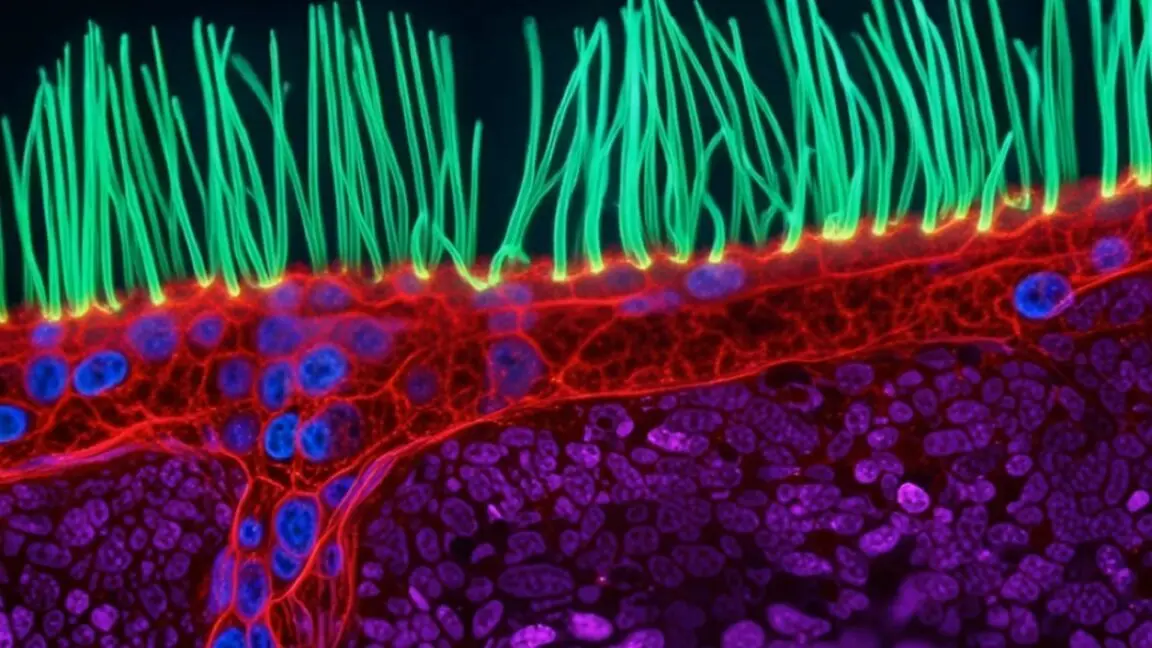
Now, a monumental shift is underway in the optical engineering and cellular biology communities. Researchers are moving past static imagery to capture the living, breathing mechanics of the cellular world. This paradigm shift was dramatically highlighted by the recent results of the prestigious Nikon Small World in Motion competition, where an innovative optical engineer claimed top honors by filming the frantic, microscopic beating of cilia—hair-like cellular structures whose malfunction leads to debilitating human respiratory and reproductive diseases.
Utilizing an advanced technique known as diffractive super-resolution microscopy, optical engineers can now observe living cells in motion over extended periods without destroying them with harsh light or toxic fluorescent dyes. This breakthrough addresses one of the most stubborn hurdles in modern biomedical research: how to record high-resolution, high-speed biological processes over sustained timeframes without inducing phototoxicity or photobleaching.
Beyond the technological triumph of the top prize, this year’s international competition showcased an astonishing array of dynamic micro-phenomena, from the synchronized division of neighboring cells to the graceful choreography of jellyfish larvae suspended in micro-droplets. This article provides a comprehensive examination of these breakthroughs, detailing the technological hurdles overcome by optical engineers, the profound implications for disease research, the chronological context of the competition, and a forward-looking analysis of where dynamic microscopy is heading next.
Detailed Chronology: The Evolution of Dynamic Micro-Observation and the 2026 Competition
To appreciate the significance of the latest advancements in microscopy, one must trace the winding historical path that brought optical engineering and cellular biology to this convergence. The journey from static plates to real-time, high-resolution video capture is a testament to decades of interdisciplinary collaboration.
Phase One: The Limits of the Static Past
For generations, microscopists faced a cruel biological paradox. To observe cellular structures with sufficient clarity, samples frequently had to be killed, fixed onto glass slides, and treated with chemical stains or fluorescent markers. While this approach illuminated intricate anatomical details, it completely obscured function. As optical engineers frequently note, viewing a fixed cell to understand a dynamic disease is akin to examining a car crash to understand how an engine works; you see the aftermath, but you miss the mechanical failure in real time.
When researchers attempted to film living specimens, they encountered severe technical roadblocks. High-speed cellular processes—such as the lashing of cilia or the rapid transport of organelles—demand rapid frame rates. Historically, achieving high resolution at high frame rates required immense amounts of illumination. However, exposing living biological samples to intense light triggers severe phototoxicity, rapidly destroying the very cellular machinery scientists intended to study. Furthermore, many fluorescent dyes used to enhance contrast cause chemical damage to living tissue over extended exposures.
Phase Two: The Rise of Diffractive Super-Resolution
The turning point arrived with the development of sophisticated light-shaping technologies. Researchers began experimenting with methods to manipulate wavefronts, utilizing spatial light modulators and digital micromirror devices to control precisely how photons interact with a biological sample.
The breakthrough crowned in the recent competition centers on diffractive super-resolution microscopy. By employing multiple light waves and a digital micromirror device to meticulously shape the illumination field, engineers found a way to bypass traditional intensity limits. Instead of flooding a specimen with a brute-force spotlight, this technique distributes light strategically, minimizing the overall photon dosage received by the living sample while maximizing the signal-to-noise ratio.
Phase Three: The 2026 Competition Showcase
The culmination of these technological leaps was vividly displayed in the latest entries to the global competition. The top prize was awarded to an optical engineer specializing in dynamic cellular imaging, who successfully filmed the relentless, rhythmic beating of living cilia.
Following this grand-prize-winning entry, the international podium reflected the diverse beauty and complexity of the microscopic realm:
- Second Place: Nguyen Nam Nhat (Vietnam), who captured an astonishing video sequence detailing the interactions and movements of a tiny roundworm alongside a single-celled organism.
- Third Place: Benedikt Pleyer (Bavaria, Germany), whose entry focused on the delicate, mesmerizing locomotion of jellyfish larvae suspended within microscopic water droplets.
- Fourth Place: Andrew Moore (Howard Hughes Medical Institute, United States), who provided unprecedented visual evidence of synchronized cell division occurring simultaneously in adjacent cells.
- Fifth Place: Patrick Hickey (Hypha Research Ltd., Edinburgh, Scotland), who documented the intricate intracellular dynamics of mitochondria and chloroplasts operating within the leaf cells of a Turtle Vine.
Supporting Context & Metrics: Overcoming the Biological and Optical Barriers
To fully grasp the magnitude of these achievements, one must examine the specific physical and biological constraints that researchers must navigate. The cellular environment is a crowded, high-speed, three-dimensional theater operating at spatial scales measured in nanometers and temporal scales measured in milliseconds.
The Cilia Conundrum
Cilia are microscopic, hair-like structures that project from the surface of many eukaryotic cells. In the human body, they play critical roles in clearing mucus and trapped pathogens from the respiratory tract, propelling ova through the fallopian tubes, and facilitating cerebrospinal fluid flow in the brain.
When cilia fail to beat in a coordinated, rhythmic fashion—often due to genetic mutations affecting their internal motor proteins, such as dynein—the consequences are devastating. Conditions like Primary Ciliary Dyskinesia (PCD) lead to chronic respiratory infections, bronchiectasis, and infertility.
Studying these conditions requires observing cilia in active motion. However, researchers face three compounding challenges:
- Diminutive Scale: Cilia are typically only a few micrometers in length and fractions of a micrometer in diameter.
- Velocity: Their beat frequency is remarkably high, often exceeding tens of beats per second.
- Fragility: They must be imaged while completely alive and functioning in a physiological buffer, ruling out fixation.
Standard advanced microscopy techniques—such as confocal laser scanning or total internal reflection fluorescence (TIRF) microscopy—often fall short. Confocal microscopy can induce significant photobleaching over time, while traditional widefield approaches lack the optical sectioning needed to resolve individual cilia amidst the visual clutter of surrounding tissue.
The Engineering Solution: Light Shaping and Digital Micromirrors
The winning methodology sidesteps these limitations by fundamentally rethinking how light is delivered to the specimen. By integrating digital micromirror devices (DMDs)—arrays comprising hundreds of thousands of microscopic, individually addressable aluminum mirrors—engineers can sculpt the spatial distribution of light dynamically.
| Microscopy Challenge | Traditional Approach | Diffractive Super-Resolution Approach |
|---|---|---|
| Illumination Intensity | High-intensity flood lighting | Precisely shaped, low-dose light waves |
| Sample Viability | Rapid phototoxicity and cell death | Extended viability, enabling ~10-second observation windows |
| Temporal Resolution | Grainy snapshots or motion blur | High-resolution capture of rapid cellular mechanics |
| Dye Requirements | Heavy reliance on phototoxic fluorescent dyes | Minimized reliance on chemical additives |
By optimizing light delivery, the system achieves what experts describe as the "sweet spot" of live-cell imaging: enough photons are collected to resolve sub-micrometer structures at high frame rates, yet the thermal and photochemical stress inflicted on the living tissue remains low enough to maintain normal cellular function throughout the observation window.
Official Statements and Perspectives from the Scientific Frontline
The intersection of optical engineering and cellular biology has fostered a collaborative culture where technical innovation directly serves medical discovery. The creators and judges behind these micro-cinematic achievements offer profound insights into the philosophy and future of the field.
The Imperative of Time
Highlighting the absolute necessity of moving beyond static photography, the grand-prize-winning optical engineer emphasized the temporal dimension of pathology:
"To understand this disease, you need to observe around 10 seconds of motion, not just a still image," the engineer explained, reflecting on the mechanics of ciliary disorders. "When people watch cilia beating, they immediately understand that something beautiful and important is happening inside us. I think this competition turns microscopy into a shared language."
This perspective underscores a broader educational and communicative goal. While high-resolution videos serve as indispensable diagnostic and research tools, they also possess a profound aesthetic resonance. By translating complex biophysical interactions into visually arresting narratives, these recordings bridge the gap between specialized laboratory research and public appreciation for the intricacies of life.
The Global Scope of Discovery
The international distribution of the competition’s top winners—spanning Vietnam, Germany, the United States, Scotland, and beyond—highlights the decentralized, globally collaborative nature of modern microscopy. Researchers across different continents are leveraging open-source optical designs, advanced computational processing, and novel fluorescent proteins to push the boundaries of what can be visualized.
Judges and industry veterans note that the standard of entries has risen exponentially over the past decade. What was once considered cutting-edge institutional research—such as capturing synchronized cell division or intracellular organelle transport—can now be achieved with remarkable fidelity by specialized research groups and independent laboratories worldwide.
Future Outlook: Where Dynamic Microscopy Is Headed Next
As we look toward the horizon of optical engineering and biomedical science, several transformative trends are poised to redefine the capabilities of dynamic microscopy. The convergence of machine learning, adaptive optics, and novel photonic materials promises to push spatial and temporal resolutions even further.
Artificial Intelligence and Real-Time Adaptive Optics
One of the most promising frontiers is the integration of artificial intelligence (AI) and deep-learning algorithms into microscope control systems. Biological samples are inherently heterogeneous and scattering; light passing through tissue wavefronts becomes distorted, leading to aberrations that degrade image quality.
Future microscopy systems will increasingly utilize real-time adaptive optics—guided by neural networks—to measure and correct these optical distortions dynamically. This will allow researchers to peer deeper into living tissues, such as intact organoids or embryonic models, without losing subcellular resolution. AI algorithms will also assist in denoising low-light video feeds on the fly, allowing for even gentler illumination protocols that preserve cell health over hours or days rather than mere seconds.
Correlative Multimodal Imaging
Another major trajectory is the rise of correlative multimodal imaging, where dynamic light microscopy is seamlessly integrated with other analytical modalities, such as atomic force microscopy (measuring mechanical properties) or label-free Raman spectroscopy (measuring chemical composition). By synchronizing these techniques in real time, scientists will not only watch how a cell moves or divides, but simultaneously measure the biochemical and biophysical forces driving those actions.
Democratization and Shared Languages
As optical components become more accessible and computational power continues to surge, the tools of high-end dynamic microscopy are gradually trickling down from elite national laboratories to university classrooms and regional research centers. This democratization accelerates the pace of discovery, ensuring that brilliant minds from diverse geographic and economic backgrounds can contribute to our understanding of cellular mechanics.
Ultimately, the milestones achieved by this year’s competition winners remind us that the universe within us is as vast, complex, and awe-inspiring as the cosmos above. By refining our ability to observe life in motion, we move closer to curing the diseases that disrupt it, transforming abstract biological challenges into tangible, solvable mechanics.